This page discusses scanning electron microscopy on uncoated insect samples.
Article:
Tardi NJ, Cook ME, Edwards KA. 2012. Rapid phenotypic analysis of uncoated Drosophila samples with low-vacuum scanning electron microscopy. Fly (Austin) 6:184-192. http://dx.doi.org/10.4161/fly.20525. [Cover article]
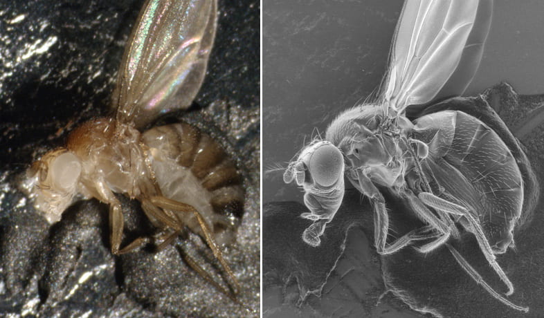
Scanning electron microscopy (SEM) is traditionally performed on metal coated samples. We refined the Low Vacuum SEM technique to allow the investigator to obtain high quality images just by placing an uncoated insect on a stub and scanning it; this cuts down the sample prep time to about 5 minutes. Our method permits SEM to be used for large-scale imaging projects such as genetic screens.
The work is based on the FEI Quanta 450 Environmental SEM, but the principles should apply to other SEM models.
See our article using one of these links:
https://www.landesbioscience.com/journals/fly/2012FLY0006R.pdf
https://www.landesbioscience.com/journals/fly/article/20525/
http://www.ncbi.nlm.nih.gov/pubmed/22722327
We also provide all the raw TIFF data from the figures. This can help you see subtle differences in image quality from varying the SEM imaging parameters.
The data are available in this sharepoint library:
https://cas.sharepoint.illinoisstate.edu/Biology/microscopy/default.aspx
If you have any trouble obtaining the images please let us know.
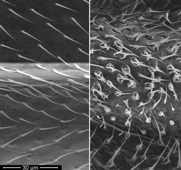
Here is an example of mutant phenotype documentation, wild type vs. mutant wing vein and wing hairs:
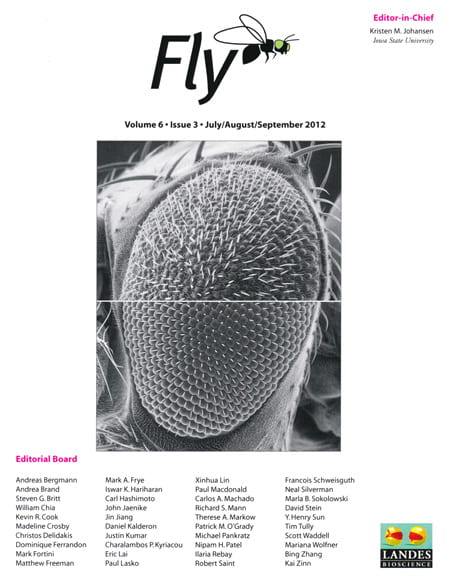
Here is our cover image from the issue: